B 1/7 Mahanagar Extension ( Opp. Sahara India Centre ), Kapoorthala, Lucknow - 226006
Mon - Sat: 10:00am to 8:30pm
Your eye doctor just told you that you need something called a Fundus Fluorescein Angiography. You nodded, said yes, and walked out wondering — what exactly is that? Is it a scan? Is it a surgery? Does it hurt? Why do I need it when I already had a full eye examination?
You are not the only one asking these questions. At Susanjeevani Hospital, the best eye hospital in Lucknow, we recommend Fundus Fluorescein Angiography — commonly called FFA — to patients every week.
They come from Gomti Nagar, Hazratganj, Indira Nagar, Alambagh, Charbagh, Mahanagar, Aliganj, Rajajipuram, and even from neighbouring districts like Barabanki, Unnao, Sitapur, and Hardoi — specifically for this investigation, because very few eye hospitals in Lucknow and the surrounding region offer it in-house.
Every time we recommend this test, we sit down and explain exactly what it is, why it is needed, and what will happen — because a patient who understands their investigation worries less, cooperates better, and gets far more from their consultation.
This blog gives you that complete explanation — in plain, simple language that anyone can understand, whether or not you have a medical background.
What Is Fundus Fluorescein Angiography?
Let us break the name into simple parts.
Fundus means the back of the eye — the retina, the macula, the optic disc, and the network of blood vessels running across them. It comes from the Latin word for "bottom" — and the retina is literally the back wall of your eye.
Fluorescein is a safe, bright orange dye that glows vividly under a specific wavelength of blue light. It is the dye used in this test.
Angiography means imaging of blood vessels — from the Greek words for "vessel" and "to write or record."
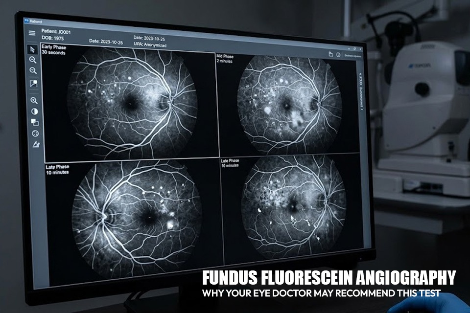
Put it all together: Fundus Fluorescein Angiography is a test where a special dye is injected into a vein in your arm, and as that dye travels through the blood vessels at the back of your eye, a special camera takes rapid sequential photographs of those vessels.
The result is a detailed, dynamic map of the blood supply to your retina — showing exactly where blood flow is normal, where it is blocked, where vessels are leaking, and where abnormal new vessels have grown.
No other investigation gives your eye doctor this level of real-time information about your retinal circulation. Not a standard eye examination, not an OCT scan, not a simple fundus photograph.
FFA is in a class of its own for assessing the vascular health of your retina.
Why Is Fundus Fluorescein Angiography Done? — Conditions It Helps Diagnose and Monitor
Your eye specialist in Lucknow will recommend an FFA when they need detailed information about the blood vessels in your retina that a clinical examination alone cannot provide. Here are the most common reasons:
1. Diabetic Retinopathy — The Most Common Reason for FFA in Lucknow
Diabetes is one of the most widespread health conditions in Lucknow and across Uttar Pradesh. Areas like Gomti Nagar, Indira Nagar, Aliganj, Mahanagar, Vikas Nagar, and the older localities of Hazratganj and Aminabad have large populations of diabetic patients — many of whom are unaware that their eyes are being silently damaged by years of high blood sugar.
Diabetic retinopathy is one of the leading causes of blindness in India. Diabetes damages the tiny blood vessels in the retina over time, causing them to leak, block, or grow abnormally.
An FFA in diabetic retinopathy shows your eye doctor precisely which blood vessels are leaking fluid into the retina, which areas of the retina are not receiving adequate blood supply — called retinal ischaemia — and whether dangerous new blood vessels have started growing.
This information directly guides treatment decisions — whether laser treatment for diabetic retinopathy, anti-VEGF injections, or advanced vitreoretinal surgery is needed, and exactly where laser spots need to be applied for maximum effectiveness.
2. Diabetic Macular Oedema — Pinpointing Exactly Where Fluid Is Leaking
Diabetic macular oedema is swelling of the macula — the central part of the retina responsible for sharp, detailed central vision used for reading, recognising faces, and watching television.
When the macula swells due to leaking retinal blood vessels, central vision blurs progressively.
This condition is particularly common among diabetic patients in Lucknow who have had poorly controlled blood sugar for many years — a pattern we see regularly in patients referred from Charbagh, Rajajipuram, Alambagh, and the older, more densely populated areas of the city where access to regular diabetic eye checkups has historically been limited.
An FFA in macular oedema identifies the exact leaking vessels causing the swelling — information essential for focal laser treatment and anti-VEGF injection planning.
It complements the OCT scan — which shows how much fluid is present — by revealing the source of the leakage.
Read our guides on how diabetes affects your eyes and diabetic eye disease to understand how macular oedema fits into the full spectrum of diabetic eye complications.
3. Retinal Vein Occlusion — Mapping the Blocked Vein and Its Impact
A retinal vein occlusion occurs when a vein draining blood from the retina becomes blocked — either the main central vein or one of its branches.
This causes a backup of blood and fluid, leading to haemorrhages across the retina and swelling of the macula.
The result is sudden, painless blurring or loss of vision in one eye — often noticed first thing in the morning.
Retinal vein occlusion is more common in patients with high blood pressure, diabetes, and high cholesterol — all conditions that are highly prevalent among middle-aged and older residents of Lucknow, particularly in areas like Gomti Nagar, Mahanagar, and Aliganj.
Many patients who come to our eye hospital in Lucknow from Sultanpur Road, Faizabad Road, and Hardoi Road corridors present with this condition after noticing sudden vision changes.
An FFA maps the full extent of the affected retina, identifies areas of ischaemia at risk of triggering dangerous new vessel growth, and guides decisions on laser treatment versus anti-VEGF injection therapy.
4. Wet Age-Related Macular Degeneration — Detecting Abnormal Vessel Growth Under the Macula
Wet age-related macular degeneration (wet AMD) is caused by the growth of abnormal new blood vessels beneath the macula.
These vessels bleed and leak fluid, causing rapid and severe central vision loss if untreated.
It most commonly affects people above 60 years of age.
With Lucknow's ageing population — particularly in established residential areas like Mahanagar, Indira Nagar, Sector C, and Gomti Nagar — wet AMD is increasingly seen at our eye clinic in Lucknow.
Families from Barabanki, Unnao, Rae Bareli, and Sitapur also bring elderly relatives to Susanjeevani Hospital specifically for advanced retinal evaluation because these investigations are not easily available closer to home.
An FFA confirms the diagnosis of wet AMD, identifies the location and type of abnormal vessels, and guides anti-VEGF injection treatment planning.
5. Retinal Artery Occlusion — Emergency Assessment of Blood Flow
A retinal artery occlusion is a medical emergency — essentially a stroke of the eye.
The artery supplying blood to part or all of the retina becomes blocked, causing sudden, severe, painless vision loss.
Time is critical — the faster treatment begins, the better the chance of preserving any remaining vision.
Patients from across Lucknow — whether from the busy commercial areas near Hazratganj and Aminabad or from quieter residential neighbourhoods in Gomti Nagar Extension and Sultanpur Road — who experience sudden vision loss in one eye should reach the best eye hospital in Lucknow as quickly as possible.
An FFA in this situation maps the extent of retinal tissue that has lost blood supply and helps guide urgent management decisions.
6. Glaucoma — Assessing Optic Disc Blood Supply
In certain types of glaucoma — particularly normal tension glaucoma where optic nerve damage occurs despite normal eye pressure — an FFA helps assess the blood supply to the optic nerve head. Poor blood flow to the optic disc is a significant contributing factor in some patients, and FFA provides direct evidence of this that guides treatment. Read our detailed glaucoma awareness and treatment guide to understand the broader context of glaucoma management.
7. Eye Floaters and Flashes — Ruling Out Serious Retinal Vascular Causes
While most floaters are benign, some floaters and flashes of light are caused by serious retinal conditions including retinal tears, vitreous haemorrhage from abnormal vessels, or inflammatory retinal vasculitis.
When your eye doctor examines the retina and suspects a vascular or structural cause, an FFA may be recommended to investigate further.
Read our guide on eye floaters and flashes — symptoms, causes and treatment to understand when these symptoms need urgent evaluation at an eye hospital in Lucknow.
8. Retinal Vasculitis and Inflammatory Eye Conditions
Retinal vasculitis is inflammation of the retinal blood vessels — caused by various systemic conditions including tuberculosis, sarcoidosis, Behcet's disease, and autoimmune disorders.
TB-related retinal vasculitis is particularly relevant in Lucknow's clinical context given the high burden of tuberculosis in Uttar Pradesh.
FFA is the gold-standard investigation for diagnosing retinal vasculitis — it shows characteristic patterns of vessel leakage, staining, and blockage that confirm the diagnosis and guide treatment decisions.
9. Monitoring Treatment Response
FFA is not just a diagnostic tool — it is also used to monitor how the retina is responding to treatment.
After a course of anti-VEGF injections or laser treatment, a repeat FFA shows whether leaking vessels have closed, whether ischaemic areas have been adequately treated, and whether further treatment is needed.
It gives your vitreoretinal specialist at our eye hospital in Lucknow objective, real-time evidence of treatment effectiveness — not just a clinical impression.
How Is Fundus Fluorescein Angiography Done? — A Complete Step-by-Step Guide
Step 1 — Pupil Dilation
Dilating eye drops are applied to both eyes. These take approximately 20 to 30 minutes to work fully.
During this time and for 3 to 4 hours after, your near vision will be blurry and your eyes will be sensitive to bright light — Lucknow's intense outdoor light, particularly during the April to June summer months, can be quite uncomfortable during this period.
Bringing sunglasses is strongly recommended.
Step 2 — Baseline Fundus Photographs
Before the dye is injected, baseline photographs of your retina are taken using the fundus camera.
These serve as a reference point to compare with the angiography images.
Step 3 — The Fluorescein Dye Injection
A small needle is inserted into a vein in your arm or hand — similar to a routine blood test.
The fluorescein dye is then injected rapidly. It takes about 5 to 10 seconds to inject and reaches the blood vessels of your retina within approximately 10 to 15 seconds.
You may experience a warm flush or a brief sensation of nausea for a few seconds immediately after the injection — this is normal and passes within a minute.
Tell the technician or doctor immediately if you feel unwell.
Step 4 — Rapid Sequential Photography
As soon as the dye is injected, the fundus camera begins taking rapid sequential photographs — several per second in the first few minutes, then at intervals over the next 5 to 10 minutes.
You will need to keep your eye open and focused on a target light during this time.
The camera emits a bright blue light during each photograph — this is the light that makes the fluorescein dye glow and makes the blood vessels visible.
The total photography time is typically 10 to 15 minutes.
Late-phase photographs may be taken at 5 to 10 minutes after injection to look for late leakage patterns.
Step 5 — After the Test
Once all photographs are taken, the needle is removed and a small bandage is applied.
Your skin and eyes may appear slightly yellowish for a few hours — harmless and temporary.
Your urine will be bright orange or yellow for 24 to 36 hours — this is the dye being excreted by your kidneys and is completely normal.
Drink plenty of water to help clear it faster.
Does the FFA Test Hurt?
No. The only discomfort is the needle insertion for the dye injection — similar to a standard blood test.
The bright camera light during photography may cause temporary dazzling but no pain.
The most noticeable effect after the test is blurry vision from pupil dilation, which lasts 3 to 4 hours.
Patients who travel to our eye hospital in Lucknow from distant areas like Chinhat, Gomti Nagar Extension, Sultanpur Road, or from outside Lucknow such as Barabanki and Unnao should factor this into their travel planning — arrange a companion to accompany you for the journey home as your vision will not be safe for driving.
FFA vs OCT Scan — What Is the Difference and Why Might You Need Both?
An OCT scan and an FFA are two different and complementary investigations — not alternatives to each other.
An OCT scan is a non-invasive scan that produces cross-sectional images of the retinal layers.
It shows the thickness of the retina, the presence and amount of fluid within retinal layers, the condition of the macula, and the structure of the optic nerve fibre layer.
It does not show blood vessel function or circulation.
An FFA is a dynamic investigation that shows blood vessel function — where vessels are leaking, blocked, or growing abnormally.
In many retinal conditions — particularly diabetic retinopathy and macular oedema — your eye specialist at Susanjeevani Hospital will use both: the OCT to measure fluid and retinal thickness, and the FFA to map vascular activity and guide laser treatment placement.
Together they give a complete picture that neither test alone can provide.
Is Fundus Fluorescein Angiography Safe? — Risks and Precautions
FFA is a very safe investigation with a long, well-established track record in ophthalmology worldwide.
Serious complications are rare.
Common, Mild Side Effects
- Temporary nausea or flushing immediately after dye injection — passes within a minute
- Yellow discolouration of skin for a few hours — harmless and temporary
- Bright orange or yellow urine for 24 to 36 hours — normal and expected
- Temporary blurry vision from pupil dilation — resolves in 3 to 4 hours
- Brief dazzling or afterimages from the camera's bright light
Uncommon Side Effects
- Extravasation — leakage of dye outside the vein at the injection site, causing localised burning and temporary skin staining. Not dangerous but can be uncomfortable.
- Vomiting — rare, occurs within the first few minutes of dye injection in a small number of patients.
Rare but Serious Reactions
Severe allergic reactions to fluorescein dye are rare — occurring in approximately 1 in 200,000 procedures.
At Susanjeevani Hospital, our eye hospital in Lucknow is fully equipped with emergency medications and trained staff to manage any allergic reaction promptly and safely.
Who Should Not Have an FFA?
- Pregnant women — fluorescein dye is not recommended during pregnancy
- Patients with known severe allergy to fluorescein dye
- Patients with severe kidney disease — extra caution required as the dye is cleared by the kidneys
Always inform your eye doctor in Lucknow about all medical conditions, allergies, and current medications before the test.
How to Prepare for Your FFA at Susanjeevani Hospital — Best Eye Hospital in Lucknow
- Do not wear contact lenses on the day of the test. Remove them the night before and wear glasses instead. Contact lenses need to be removed before dilation drops are applied.
- Arrange for someone to accompany you. Your pupils will be dilated for 3 to 4 hours after the test. Do not drive.
- Whether you are coming from nearby Mahanagar or travelling from Faizabad Road, Hardoi Road, or Barabanki, please have a companion or arrange a cab for the return journey.
- Bring sunglasses. Lucknow's bright sunlight — especially during summer months — combined with dilated pupils makes outdoor light genuinely uncomfortable. Sunglasses are essential for the journey home.
- Eat and drink normally before the test. No fasting required. Take your regular medications including diabetes and blood pressure drugs.
- Stay well hydrated. Adequate water intake before the test helps the dye inject smoothly and clears from your system faster after.
- This is particularly important during Lucknow's hot summer months when dehydration is common.
- Inform your doctor of all allergies — particularly any known reaction to dyes, injectable medications, or iodine.
- Wear clothing with sleeves that roll up easily for the arm injection.
- Plan your travel time carefully. Patients coming from Gomti Nagar Extension, Sultanpur Road, Chinhat, or from outside Lucknow like Sitapur, Hardoi, or Rae Bareli should arrive well before their appointment time to account for Lucknow's traffic — particularly the congestion near Charbagh, Hazratganj crossing, and the Mahanagar-Kapoorthala stretch during peak hours.
- If this is your first visit to our eye hospital in Lucknow, reading our complete first-time patient guide to preparing for your eye consultation at Susanjeevani Hospital will ensure you are fully prepared for the entire visit.
Fundus Fluorescein Angiography at Susanjeevani Hospital — The Best Eye Hospital in Lucknow
Susanjeevani Hospital, located at Kapoorthala in Lucknow, is one of a very small number of eye hospitals in Lucknow with a fully equipped, in-house Fundus Fluorescein Angiography facility.
This means that when your eye specialist recommends an FFA, you do not need to travel to another centre, wait days for a referral appointment, or travel to Lucknow's major government hospitals at Chowk or Gomti Nagar just for this investigation.
It is performed right here, reviewed immediately by our vitreoretinal specialist, and the results directly inform your treatment plan on the same visit.
Patients from across Lucknow — whether from the newer IT and residential hubs like Vibhuti Khand, Gomti Nagar, and Shaheed Path corridor, or from older established localities like Aminabad, Charbagh, Naka Hindola, and Rajajipuram — as well as patients travelling from Barabanki, Unnao, Sitapur, Hardoi, Rae Bareli, and Lakhimpur Kheri choose Susanjeevani Hospital for their retinal investigations because of the quality of our in-house diagnostic facilities and the expertise of our vitreoretinal specialist.
Dr. Mohit Khemchandani, our senior ophthalmologist and vitreoretinal specialist, has extensive experience interpreting FFA images across the full spectrum of retinal conditions — from diabetic retinopathy and macular oedema to retinal vascular occlusions, wet AMD, and retinal vasculitis.
The combination of in-house FFA with our OCT scanning, anti-VEGF injection therapy, and laser treatment for diabetic retinopathy means Susanjeevani Hospital provides a complete, integrated retinal care pathway — from investigation to treatment to long-term follow-up — entirely under one roof.
Visit our Fundus Fluorescein Angiography treatment page for further details about this investigation at our hospital.
OPD Timings: Monday to Saturday, 10:00 AM to 8:30 PM
Phone: +91-8400868388
Address: B 1/7 Mahanagar Extension, opposite Sahara India Centre, Kapoorthala, Lucknow — 226006
Easily accessible from Gomti Nagar (15 minutes), Hazratganj (20 minutes), Indira Nagar (20 minutes), Alambagh (25 minutes), Charbagh Railway Station (25 minutes), and Amausi Airport (30 minutes).